Home
/ Tendon Diagram Foot : Anatomy Of The Foot Ankle Everything You Need To Know Dr Nabil Ebraheim Youtube - A foot tendon tear happens when one of the tendons in the foot is damaged from sudden injury or overuse.
Tendon Diagram Foot : Anatomy Of The Foot Ankle Everything You Need To Know Dr Nabil Ebraheim Youtube - A foot tendon tear happens when one of the tendons in the foot is damaged from sudden injury or overuse.
Tendon Diagram Foot : Anatomy Of The Foot Ankle Everything You Need To Know Dr Nabil Ebraheim Youtube - A foot tendon tear happens when one of the tendons in the foot is damaged from sudden injury or overuse.. Tendons in foot diagram (page 1) muscles that lift the arches of the feet foot tendons and ligaments diagram these pictures of this page are about:tendons in foot. One of the main ligaments in the foot is the plantar fascia, which forms the arch on the sole of the foot. Knee diagram tendons was posted in may 29, 2015 at 4:57 pm. Collection by prudence natasha jones. The achilles tendon is also called the calcaneal tendon.
The most common cause of tendonitis is overuse, which means a tendon is overly stretched and possibly experiencing a small degree of pulling apart or tearing.this occurs when there is an increase in activity, which can include anything from walking to participating in competitive sports. The big toe has a strong and large tendon that controls flexion and extension. Muscles and tendons of the dorsum of foot download scientific. Originates from the lower part of the fibula and attaches to the outer side of the midfoot The ligaments of the foot and ankle can be divided into groups including:
Atlas And Text Book Of Human Anatomy Anatomy Atlases The Ifcscles Of The Foot 229 The Synovial Sheaths Of The Foot Like The Tendons Of Ihc Muscles Of The Forearm The from c8.alamy.com Problems such as flat feet or high arches can create muscular. A tendon is band of tissue made up of many fibers. Tendon diagram of calf and knee. There are a whole range of structures e.g. Common ossicles of the foot some feet contain accessory ossicles or accessory bones (figure 9). Muscles and tendons of the dorsum of foot download scientific. Bones, muscles, tendons and nerves which will each give slightly different foot pain symptoms. 9 photos of the foot tendons and ligaments diagram.
Swelling and bruising will occur at the site of injury.
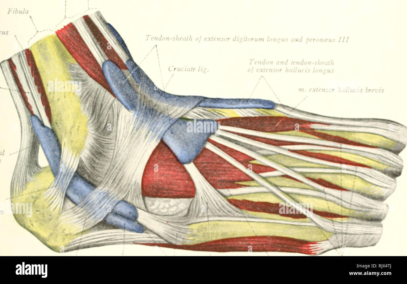
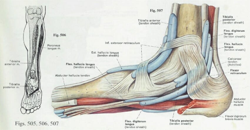
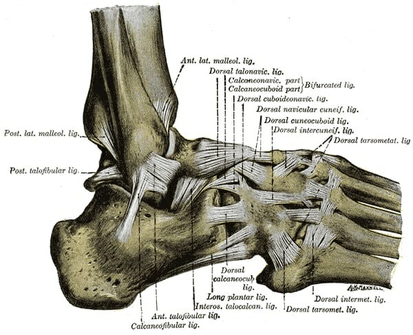
9 photos of the foot tendons and ligaments diagram. There are a whole range of structures e.g. Other tendons help to control the movements of the toes. Ligaments stabilizing the ankle joint. Other important tendons in the foot include the tibialis posterior (posterior tibial tendon), which attaches the calf muscle to the bones on the inside of the foot and supports the arch of the foot, and the tibialis anterior (anterior tibial tendon), which runs from the outer tibia to the first metatarsal and surfaces of the median cuneiform tarsal, which allows for dorsiflexion—bringing the. Pain and tenderness are concentrated on the top, bottom or the sides of your foot near the arch. A tendon is band of tissue made up of many fibers. Anatomical foot diagram catalogue of schemas. Foot anatomy diagram, foot joint diagram, foot sprain diagram, foot tendons and ligaments pain, leg this diagram depicts knee diagram tendons. Tendon diagram / a patient s guide to foot anatomy 2020 orthonorcal los gatos capitola morgan hill watsonville ca : Ligaments hold bones together and stabilize joints. Did you know that the tendon sheaths of the foot prevent the tendon from adhering to the overlying kim bengochea, regis university, denver. Tendon diagram of calf and knee.
Diagram of foot tendons written by jupiterz friday, march 23, 2018 add comment edit. Blood supply to the foot foot ankle orthobullets. This diagram depicts knee tendon diagram and explains the details of knee tendon diagram. This may be caused by sudden trauma, such as rolling the foot, which causes the ligaments to pull away from the bone. abnormal foot structure:
Foot Anatomy Bones Ligaments Muscles Tendons Arches And Skin from biologydictionary.net Ligaments are strong connective tissue composed of fibrous tissues. Originates from the upper part of the fibula, passes underneath the foot and attaches by the medial foot arch peroneus brevis: Muscles and tendons of the dorsum of foot download scientific. Did you know that the tendon sheaths of the foot prevent the tendon from adhering to the overlying kim bengochea, regis university, denver. The muscles, tendons and ligaments. Human feet are mostly subjected to injury due to their complicated structure. The tendons are thick bands that connect muscles to bones. Hi, for the last week or so, i get this sudden sharp and pulsating pain in my foot, the inner arch area (left feet).i looked at a foot muscle diagram to identify the area and it looks like the abductor hallucis area where the pain is located.
Ligaments stabilizing the ankle joint.
The foot diagram has a complex structure made up of bones, ligaments, muscles, and tendons. Originates from the upper part of the fibula, passes underneath the foot and attaches by the medial foot arch peroneus brevis: Foot anatomy diagram, foot joint diagram, foot sprain diagram, foot tendons and ligaments pain, leg tendon diagram, peroneal tendonitis, foot, foot anatomy diagram, foot joint diagram, foot sprain diagram, foot tendons and ligaments pain, leg tendon diagram, peroneal tendonitis. Extensor tendinitis happens when the tendons on top of your foot become inflamed. It is made up of bones, muscles, tendons, ligaments and 100 other which are designed o allow the foot to balance the body on two legs. The foot has a number of tendons. Originates from the lower part of the fibula and attaches to the outer side of the midfoot A tendon is a band of tissue that connects a muscle to a bone. Other tendons help to control the movements of the toes. Human feet are mostly subjected to injury due to their complicated structure. Drawing of the lower leg including the ankle and foot bones. It is a band of fibrous connective tissues. abnormal foot structure:
The tendons are thick bands that connect muscles to bones. This may be caused by sudden trauma, such as rolling the foot, which causes the ligaments to pull away from the bone. There are a whole range of structures e.g. Diagram of foot tendons written by jupiterz friday, march 23, 2018 add comment edit. Ligaments hold bones together and stabilize joints.
Foot Anatomy Bones Ligaments Muscles Tendons Arches And Skin from biologydictionary.net A major tendon in the foot is the achilles tendon, which is the largest tendon in the body. Tearing any of the tendons of the foot can be very painful. They connect bones to other bones, and are extremely important in stabilizing joints. Foot anatomy diagram, foot joint diagram, foot sprain diagram, foot tendons and ligaments pain, leg this diagram depicts knee diagram tendons. Other tendons help to control the movements of the toes. The foot has a number of tendons. The achilles tendon is also called the calcaneal tendon. A foot pain diagram is a great tool to help you work out what is causing your ankle and foot pain.
Download scientific diagram | tendon structure and composition.
The muscles are located mainly in the sole of the foot and divided into a central (medial) group and a group on either side (lateral). A foot pain diagram is a great tool to help you work out what is causing your ankle and foot pain. Ligaments hold bones together and stabilize joints. It is a band of fibrous connective tissues. The foot has a number of tendons. The muscles, tendons and ligaments. Drawing of the lower leg including the ankle and foot bones. These tendons help your extensor muscles pull your foot upwards, which is necessary for walking. Tendons in foot diagram (page 1) muscles that lift the arches of the feet foot tendons and ligaments diagram these pictures of this page are about:tendons in foot. Foot anatomy diagram, foot joint diagram, foot sprain diagram, foot tendons and ligaments pain, leg this diagram depicts knee diagram tendons. Anatomical foot diagram catalogue of schemas. Here you can see the tendons that extend down the top of your foot toward your toes, allowing you to curl your toes upward if need be. Ligaments stabilizing the ankle joint.
9 photos of the foot tendons and ligaments diagram tendon diagram. One of the main ligaments in the foot is the plantar fascia, which forms the arch on the sole of the foot.